Epithelial Cell Diagram Labeled
Epithelium ciliated columnar cells epithelial function histology quizlet elongated nuclei Intestinal epithelial cell royalty-free stock image Epithelial epithelium anatomical squamous vectormine tissues
Histology Image: Membranous epithelium
What is epithelial tissue different types of structure location and Secretion epithelial modes epithelium columnar glands exocrine glandular correctly methods merocrine apocrine physiology Epithelial epithelium class tissues ciliated biology describe pseudostratified cilia comprises layer kind
Epithelial tissue types characteristics classification epithelium notes biology
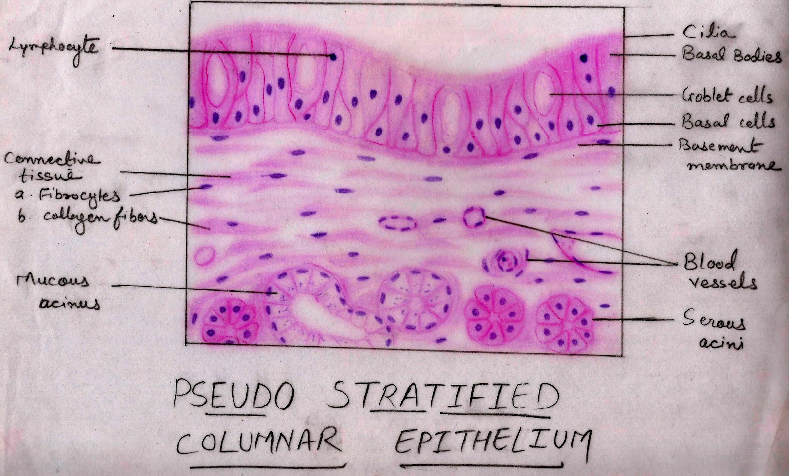
Epithelial cell intestinal microvilli illustration illustation detailed brightEpithelium histology cells layer nuclei membranous columnar stratified pseudo single two variable shape level height Describe various types of epithelial tissues with the help of labeledEpithelial tissue · anatomy and physiology.
Epithelium epithelia histology glands cells goblet cell columnar pseudostratified tissue simple unicellular exocrine do diagram stomach subtypes fully membrane partsCells epithelial epithelium tissue cell goblet stratified lining human anatomy columnar intestine micrograph small surface sample simple body tissues single Epithelial tissue cells tissues shape cell classified cover line surfaces epithelium classification simple arrangement innervated medicinebtg other sizesTissue epithelium stratified epithelial nonkeratinized squamous function cuboidal structure location cells keratinized columnar simple non where types different found esophagus.

Transitional epithelium tissue functions epithelial tissues found lesson study ck foundation slide
Epithelial tissues epithelium columnar layer lie consists nuclei| schematic diagram of jnk signaling in the tubular epithelial cell Epithelia: the histology guideImage gallery transitional epithelium.
Histology image: membranous epitheliumCiliated columnar epithelium Describe various types of epithelial tissues with the help of labeledEpithelial tubular jnk signaling response.

34 correctly label the following areas on a slide of simple columnar
Epithelial cells vector illustration – vectormineCells in the epithelial tissue Epithelial tissue: characteristics and classification scheme and types.
.


Describe various types of epithelial tissues with the help of labeled

Describe various types of epithelial tissues with the help of labeled

34 Correctly Label The Following Areas On A Slide Of Simple Columnar

Ciliated Columnar Epithelium

Epithelial Tissue · Anatomy and Physiology

Epithelial cells vector illustration – VectorMine

| Schematic diagram of JNK signaling in the tubular epithelial cell

Image Gallery transitional epithelium
Histology Image: Membranous epithelium